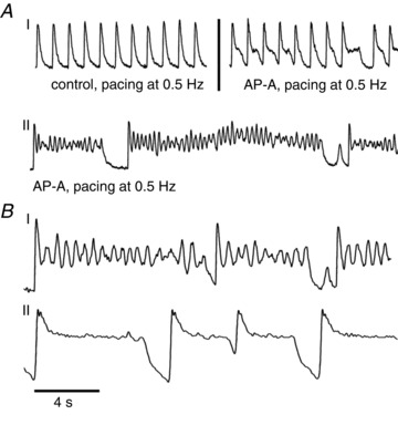
Figure 2. The effect of AP-A on isolated and connected neonatal rat ventricular myocytes.

A, panel I (left) perfused with normal Tyrode, paced at 0.5 Hz; (right) AP-A 4 nm perfusion, paced at 0.5 Hz; panel II) AP-A 4 nm perfusion, paced at 0.5 Hz. B, comparison between isolated cardiomyocyte (I) and confluent monolayer (II), paced at 0.25 Hz. AP-A, anthopleurin-A.
