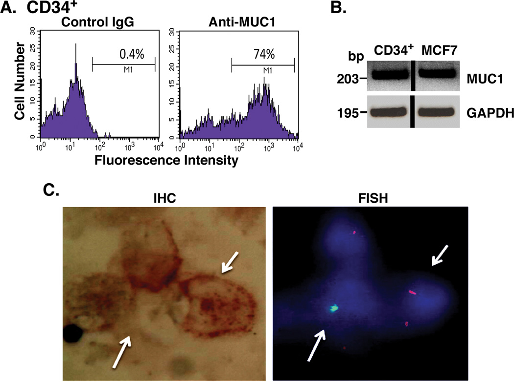
Figure 2. MUC1 expression by recipient cells in a patient following allogeneic transplantation.
CD34+/lineage− cells were isolated from the bone marrow of a female patient with AML after a sex mismatched allogeneic bone marrow transplant. A. The CD34+ cell population was incubated with a control IgG (left) and anti-MUC1 (right), and analyzed by flow cytometry. B. CD34+ cells were analyzed for MUC1 and GAPDH mRNA levels by RT-PCR. RNA from MUC1+ MCF-7 breast cancer cells was used a positive control. C. CD34+ cells were analyzed for MUC1 expression by immunohistochemical staining (left) and sex chromosomes by FISH (right) using the BioView System. Representative female recipient cells (XX; red signals) and male donor cells (XY; green signals) are highlighted with arrows.

