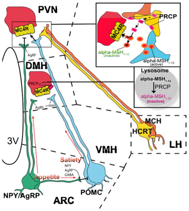
Fig. 1.
Schematic illustration showing the site of action of PRCP on α-MSH. PRCP is mainly expressed in the dorsomedial nucleus of the hypothalamus (DMH) where MC4R-expressing neurons are located and in the lateral hypothalamic (LH) hypocretin/orexin (Hcrt) and melanin-concentrating hormone (MCH) neurons. Thus, it is hypothesized that, in the DMH, PRCP degrades α-MSH at the membrane and/or intracellularly, terminating the effect of α-MSH on MC4R. From the lateral hypothalamus, Hcrt and MCH neurons project to several areas of the hypothalamus, such as the paraventricular nucleus (PVN), where α-MSH terminals strongly innervate MC4R-expressing neurons. Thus, it is hypothesized that PRCP is released from the axon terminals of Hcrt and/or MCH terminals in the PVN, degrading α-MSH extracellularly and increasing the antagonistic effect of AgRP. VMH ventromedial hypothalamic nucleus. Squares are magnifications of the corresponding zone in the figure. Dotted lines delimitate hypothalamic nuclei

