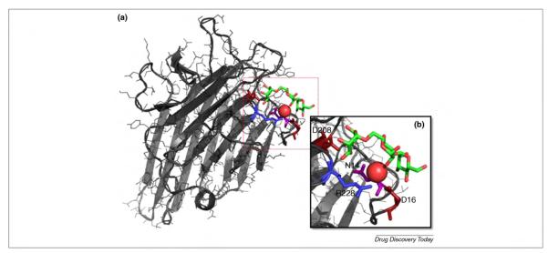
FIGURE 2.

(a) A complex of concanavalin A (ConA) with trimannoside (PDBID 1CVN [55]). (b) A close-up of ConA binding site. Key residues for carbohydrate binding and recognition are highlighted: Asp 16 and Asp 208 in red, Arg 228 in blue, and Asn 14 in purple. The trimannoside is colored according to its atom types. The oxygen of a key water molecule is also represented as a red van der Waals (vdW) sphere.
