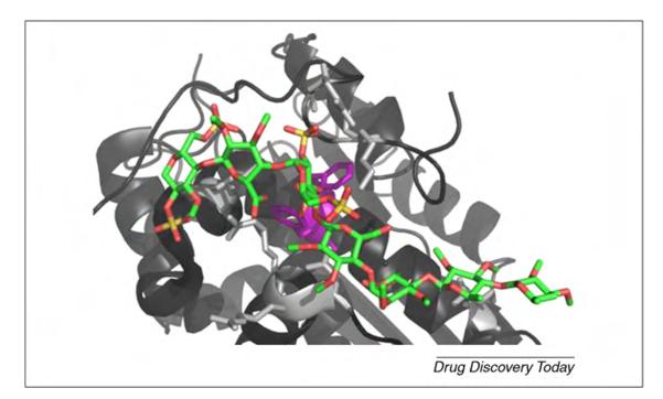
FIGURE 3.

Detail of a complex between antithrombin (AT3) and heparin (PDBID 1SR5 [135]). Heparin is colored according to its atom types, and two Phe keys for heparin binding are highlighted in purple. The Arg and Lys residues in the binding site are shown in light gray.
