
Figure 10.
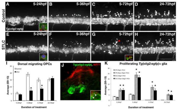
Kif11 is required for oligodendrocyte development. Lateral views of Tg(olig2:EGFP) embryos treated with either vehicle control (A–D) or STLC (E–H) at 5hpf and fixed at time points critical for oligodendrocyte development (5–24hpf, 5–36hpf, 5–72hpf, and 24–72hpf). Dorsal migrating OPCs (white asterisks) were present in control embryos older than 24hpf (B–D), which were significantly reduced in STLC treated groups (F–H, quantified in I, black asterisks p>0.05), except when treated from 24–72hpf (no statistical significance from the vehicle control, p>0.51). Irregular GFP+ fragments (G arrowhead) co-labeled with anti-Activated Caspase-3 (G, inset, red fluorescence) indicative of cell death. (J) Kif11 mRNA detection within 27hpf Tg(olig2:EGFP) embryos indicated that a small portion of olig2 cells associated with the ventricular zone colocalize with kif11 expression (J inset, arrowheads). Triple-labeled (GFP+, Ph3+, Gfap+) cells were identified in most control and treatment groups (A,E brackets; insets are maximum intensity projection of extracted Z-stack series in A,E; quantified in K), and were significantly increased in all STLC treated groups (K, asterisks). Error bars delineate the standard error of the mean. Asterisks indicate a statistical significance (t-test, two tailed, assuming equal variances, p<0.05).