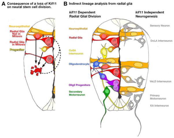
Figure 11.
Model of the role Kif11 plays in neural stem cell division and neurogenesis. (A) Illustration of one half of the neural tube depicting the consequences of a loss of kif11 to neural stem cell division. Neuroepithelial cells (orange) transition into radial glial cells (orange/red) that undergo division at the ventricular surface to generate progenitor cells (yellow) and/or additional radial glia (red). Upon loss of kif11, radial glia cells arrest in mitosis (lower red cell with monoaster spindle), which leads to increases in cell death (red apoptotic cell exhibiting cell blebbing). We propose that cell death is progressively initiated in cells at all stages of the cell cycle as opposed to cells just arrested in mitosis (three arrows). Moreover we propose that the consequential loss of cells by apoptosis will show reductions in the number of cells entering S-phase; however, we cannot rule out that negative feedback on neighboring parental stem cell populations signals a slow down in cell cycle entry (dashed inhibitory lines). (B) Examination of neuronal and glial cell fates in kif11−/− mutants provides an indirect method of lineage tracing from radial glial cells. Along the left side of this illustrated spinal cord shows the cell types that require Kif11 function, and therefore likely possess radial glial origins. Cell types that were patterned normally in the absence of functional Kif11 are illustrated along the right side of this spinal cord, and thus likely are not derived from radial glia.

