
Figure 6.
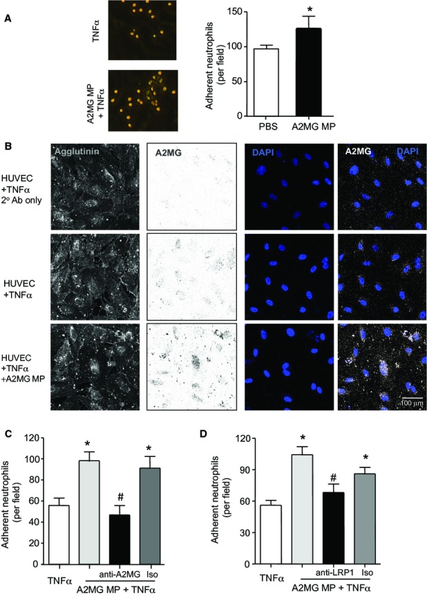
A2MG is transferred by microparticles onto endothelial cell plasma membranes and promotes neutrophil adhesion to endothelial cells. Results are mean ± s.e.m. of n = 3–4 distinct microparticle and HUVEC preparations per group (*P < 0.05, versus TNF-α; #P < 0.05 versus A2MG MP + TNF-α by Student's t-test or one way ANOVA).
A HUVEC were incubated with TNF-α (10 ng/ml) in the presence or absence of microparticles expressing elevated A2MG levels (A2MG MP) for 4 h at 37°C. Freshly prepared neutrophils were then perfused over the monolayer at 1-dyne/cm2 for 8 min and the number of adherent leukocytes quantified.
B HUVEC were stimulated with TNF-α (10 ng/ml) with or without A2MG MP (5 × 10⁴) for 4 h, these were then fixed, stained using Alexa Fluor® 633-Agglutinin (Membrane), anti-A2MG antibody (5 μg/ml) and mounted in Probing Antifade medium containing DAPI.
C A2MG MP were incubated with anti-A2MG antibody (5 μg/ml), or isotype control (Iso; 10 min, RT) prior to incubation with HUVEC monolayer (4 h, 37°C), and neutrophil perfusion.
D Neutrophils were incubated with anti-LRP1 (5 μg/ml) antibody or isotype control (Iso; 10 min, RT) prior to perfusion over endothelial cells that were incubated with A2MG MP (4 h, 37°C).