
Figure 1.
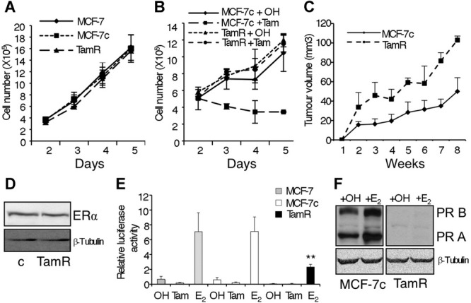
Characterization of MCF-7TamR cells.
A Proliferation assay of MCF-7 (wt), MCF-7c (control) and MCF-7TamR (tamoxifen resistant) cells (n = 3).
B Proliferation assay of MCF-7c and MCF-7TamR cells treated with ethanol (OH) or 5 × 10⁻⁷ M tamoxifen (Tam) (n = 3).
C Tumour growth curve of MCF-7c and MCF-7TamR cells implanted s.c. in athymic mice in the presence of an exogenous slow release, oestrogen implant (n = 5 mice/group).
D Western blot analysis of ERα expression in MCF-7c (c) and MCF-7TamR cells.
E MCF-7 (grey bars), MCF-7c (white bars) and MCF-7TamR (black bars) cells were transfected with a reporter plasmid containing three copies of a consensus ERE driving a luciferase reporter in the presence of the carrier ethanol (OH) or 5 × 10⁻⁷ M tamoxifen (Tam) or 10−8 M oestrogen (E2). In all transfections, β-galactosidase activity was used to control for transfection efficiency (n = 5) **p = 0.007 by t-test.
F Progesterone receptor expression in control (MCF-7c) and resistant (TamR) cells by Western blot analysis.