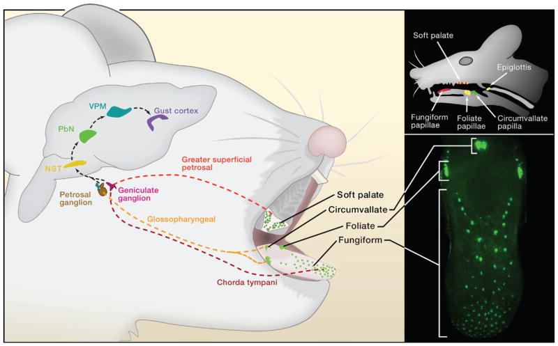
Figure 1. The Anatomy of Taste.
Taste buds are broadly distributed on the tongue and soft palate. On the tongue, taste buds are localized to three classes of papillae: In mice, the single circumvallate papilla is found at the very back of the tongue; foliate papillae are at the posterior lateral edge, and fungiform papillae are distributed over the anterior two thirds of the tongue; these three classes of paplilae can be highlighted in mice engineered to express green fluorescent protein in taste bud areas (lower right panel). The taste buds on the tongue and palate are innervated by three afferent nerves: the chorda tympani, greater superficial petrosal, and glossopharyngeal. These nerves carry taste information from the taste receptor cells to the nucleus of the solitary tract (NST) in the brain stem. From the NST, taste responses are transmitted (and processed) through the parabrachial nucleus (PbN) and the thalamus (VPM) to the primary gustatory cortex in the insula. Behavioral responses to food (and perceptions of flavor) are ultimately choreographed by the integration of gustatory information with other sensory modalities (such as olfaction, texture, etc.)

