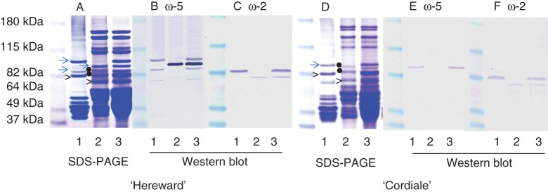
Fig. 2.

Identification of ω-gliadins in ‘Hereward’ (A–C) and ‘Cordiale’ (D–F) by SDS–PAGE (A, D) and western blot analysis with antibodies to ω-5 (B, E) and ω-2 (C, F) gliadins. The monomeric gluten proteins extracted by 50 % (v/v) propan-1-ol, reduced residual proteins (including glutenin subunits) and total proteins are shown in lanes 1, 2 and 3, respectively. The dots indicate proteins in the ω-gliadin region which did not react with either antibody and were not present in the total gluten protein fractions (Fig. 1).
