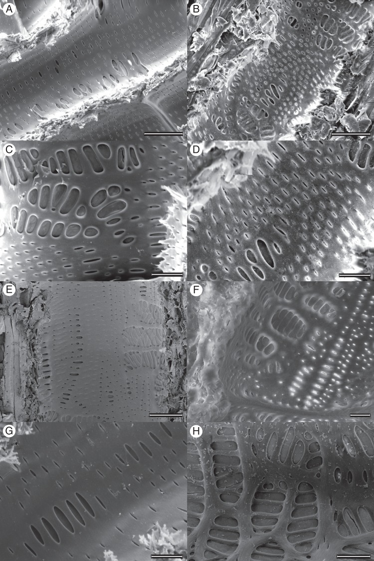
Fig. 4.
Environmental scanning electron microscopy images of Sideroxylon lanuginosum (A, C, E, G) and Quercus fusiformis (B, D, F, H) shallow (A–D) and deep (E–H) roots showing the large degree of variability in pit shape and size. Scale bars are (A, B) 50 µm, (C, D) 20 µm, (E) 50 µm and (F–H) 20 µm.

