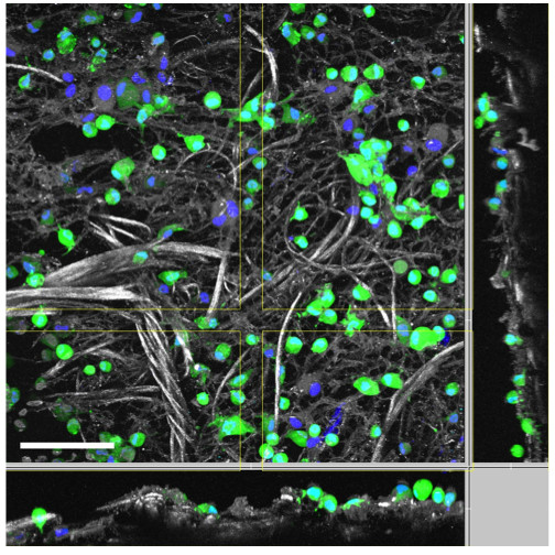
Figure 4.

Two-photon microscopy of the enriched collagen and elastin matrices. This figure shows a representative two-photon microscopy image of a collagen and elastin matrix, which was incubated with freshly isolated adipose-derived stem/stromal cells for only 1 h. The green colour indicates the cytoplasm of viable cells, which was stained using fluorescein diacetate (FDA), the blue colour indicates the cell nuclei, which were stained using Hoechst 33342, and the grey colour indicates the collagen and elastin structure (scale bar is 100 μm).
