Figure 2.
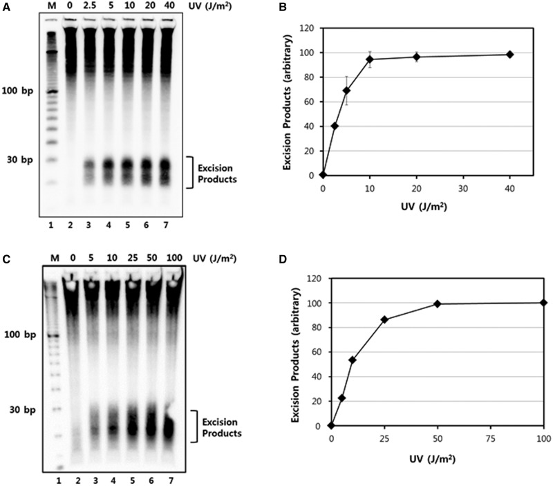
UV dose-dependent generation of excised oligonucleotides. (A) HeLa cells were treated with the indicated doses of UV-C radiation and then harvested after incubation for 30 min. Excised oligonucleotides containing UV damage were then extracted, 3′ end-labeled with biotin, separated on a 10% TBE-urea gel, transferred to a nylon membrane, and detected with streptavidin-HRP conjugate using chemiluminescence reagents. (B) Quantitative analysis of the excision repair products from three independent experiments as in A are presented as means ± standard deviation (SD). The maximum values were set to 100 and the other values are presented relative to that value. (C) A375 cells were treated with the indicated doses of UV-C radiation and then harvested 1 h later. Excised oligonucleotides were analyzed as in A, except with a 12% TBE-urea gel. (D) Quantitative analysis of the excised oligonucleotide repair products in A375 cells.

