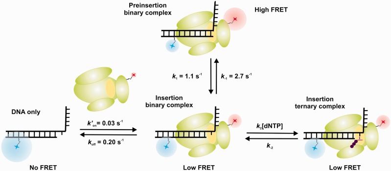
Figure 6.
Different conformations for Dpo4 binding to DNA shown schematically. Cy3 fluorophore (blue) is on the DNA template, and the Cy5 fluorophore (red) is on the polymerase. Dpo4 active site is shown in yellow. For the preinsertion binary complex, the primer terminus base is sitting in the polymerase active site at the position that an incoming nucleotide binds. The insertion binary complex has a free nucleotide binding site. In the insertion ternary complex, the incoming nucleotide is bound at the active site.

