Figure 3.
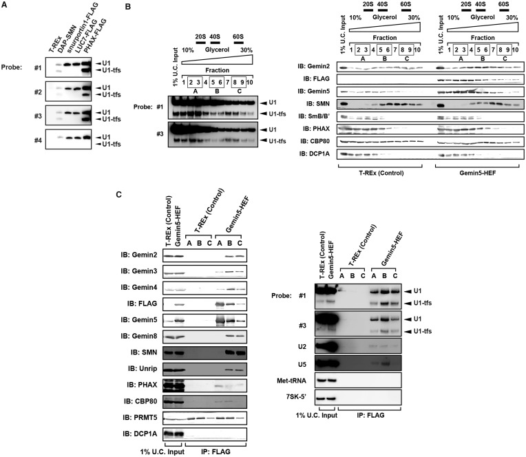
U1-tfs are formed at early steps of U1 snRNP biogenesis. (A) RNAs were pulled down using DAP-SMN, FLAG-fused PHAX, U1-70K, snurportin or LUC7 as affinity bait. RNAs were separated by denaturing urea-PAGE and visualized by northern blotting with probes #1–4. (B) Total cell extract of T-REx 293 cells (left panel) or Gemin5-HEF-expressing T-REx 293 cells (right panel) were separated into 10 fractions by glycerol gradient (10–30%) ultracentrifugation. Each fraction was analyzed by immunoblotting with antibodies against the proteins indicated. Northern blotting with DNA probe #1 or #3 showed a typical U1 RNA distribution pattern of this ultracentrifugation fractionation. Sedimentation values are indicated above the elution profile. (C) Fractions 2 and 3 (mixture A), 5 and 6 (mixture B) or 8 and 9 (mixture C) in (B) were mixed and subjected to pull-down analysis with Gemin5-HEF as affinity bait. Proteins and RNAs pulled down with Gemin5-HEF were analyzed by immunoblotting (IB) with antibodies against the proteins indicated and by northern blotting with the DNA probes indicated, respectively.

