
Figure 1.
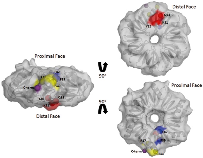
Surface representation of the WT Ec Hfq structure and the positions of the tryptophan-substituted residues. Highlighted in red are residues on the distal face that were mutated to tryptophan (Trp); in yellow are the lateral residues that were mutated to Trp, in blue the proximal face residues that were mutated to Trp and in purple the position to indicate the beginning to the C-terminal 44 residues of Ec Hf. A cartoon of the underlying hexamer is shown in grey. PDB accession number for the WT Ec Hfq structure is 1HK9 (33).