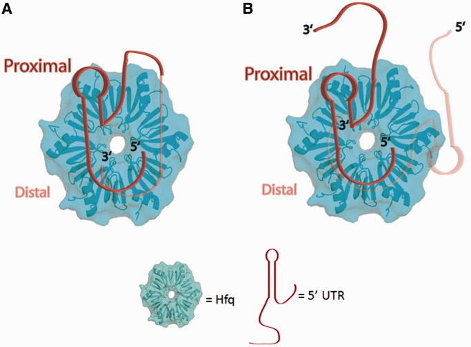
Figure 8.
Potential models for Ec Hfq binding to the 5′-UTR of hfq mRNA. (A) hfq mRNA binding such that a single mRNA uses all sets of identified binding sites to interact with both faces of Ec Hfq with the linker between the two binding sites wrapping around the outside of the Hfq hexamer. (B) Two hfq mRNA binding to the proximal and distal faces of a single Hfq hexamer. The 5′-UTR of the hfq mRNA is shown as a ribbon with its 5′ and 3′ ends labelled. The 5′ end of the mRNA contains Site A, which should wrap about the proximal pore and lead to the rim/lateral surface. The 3′ end of the mRNA contains Site B, which has a preference for the distal face and C-terminal region of Hfq. The 5′-UTR of the hfq mRNA is modelled onto a surface rendering of Hfq generated in Pymol using PDB ID: 1HK9 (33).

