
Figure 7. Zfx activates the expression of Ptpmt1 and Idh2 in leukemia cells.
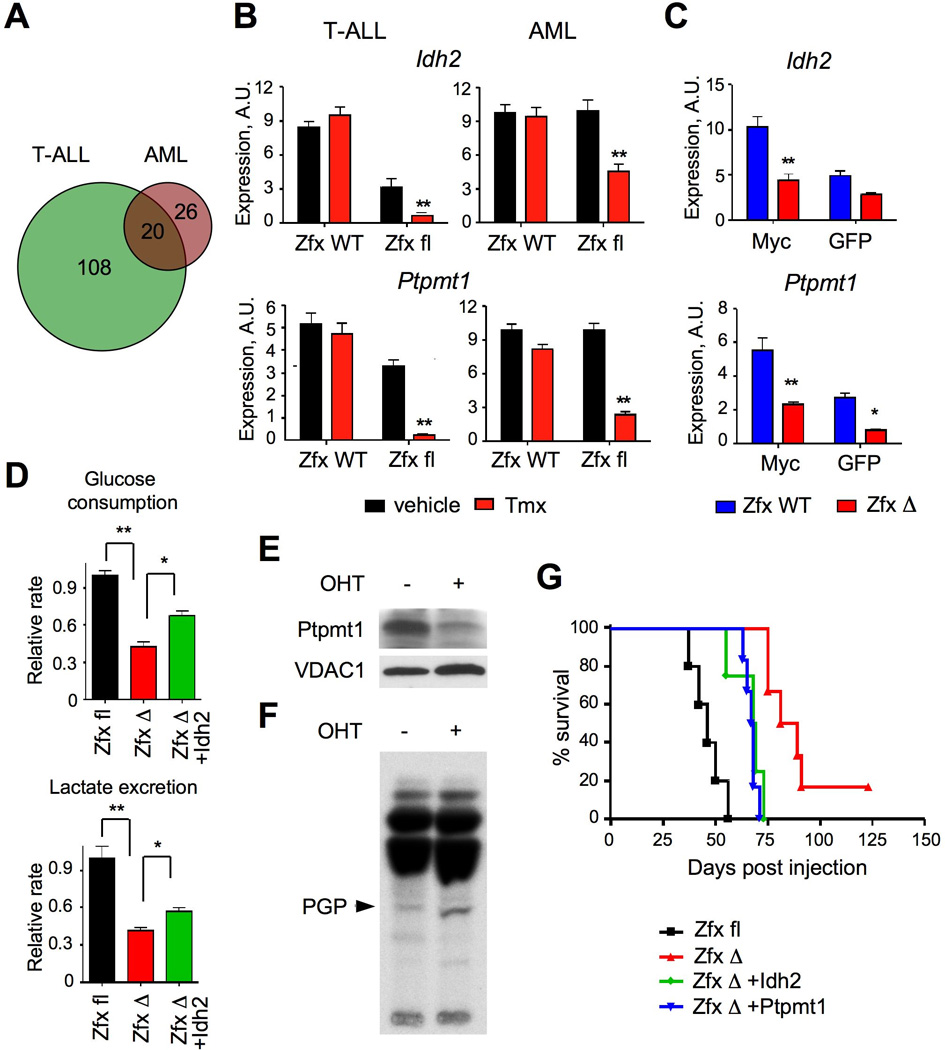
(A) The overlap between conserved direct targets of Zfx activation in T-ALL and AML.
(B) The expression of Idh2 and Ptpmt1 in murine leukemia cells after Zfx deletion. The deletion was induced by Tmx (or vehicle as a control) in the R26-CreER+ conditional (Zfxfl/y) or control (Zfxwt/y) leukemia cells. NotchIC-transformed T-ALL cells were sorted from secondary recipients 5 days after Tmx treatment; MA9-transformed primary AML were incubated with 4-OHT for 3 days. Shown are relative expression levels as determined by qRT-PCR (mean ± SEM of 5 independent lines for T-ALL and 7–8 independent lines for AML). **P<0.01.
(C) The expression of Idh2 and Ptpmt1 in Zfx-deficient progenitors during transformation by Myc. Shown are relative transcript levels determined by qRT-PCR in ZfxΔ/y or Zfxwt/y progenitors 4 days after transduction by Myc-GFP or GFP only (mean ± SEM of 6 independent cultures) * P<0.05, ** P<0.01.
(D) Effect of Zfx deletion on the glycolysis rate of murine AML cells grown in liquid culture with cytokines. MA9-transformed R26-CreER+ Zfxfl/y AML line was transduced with retroviral vectors expressing Idh2 or GFP alone. The cells were incubated with 4-OHT to induce Zfx deletion, propagated for 4 days in liquid culture and analyzed for the steady-state glucose consumption and lactate excretion rates (mean ± SEM of triplicate cultures). *P<0.05, **P<0.01.
(E–F) Effect of Zfx deletion on the expression and function of Ptpmt1. MA9-transformed R26-CreER+ Zfxfl/y AML line was incubated with 4-OHT for 5 days to induce Zfx deletion, and analyzed 4 days later by Western blotting for Ptpmt1 (panel D) and by thin layer chromatography for 32P-labeled mitochondrial lipids (panel E). The position of PGP was confirmed by parallel analysis of pure 14C-labeled PGP.
(G) Effect of Idh2 and Ptpmt1 overexpression on the growth of Zfx-deficient AML. MA9-transformed R26-CreER+ Zfxfl/y AML line was transduced with retroviral vectors expressing Idh2, Ptpmt1 or GFP alone. The cells were incubated with 4-OHT to induce Zfx deletion, and the resulting ZfxΔ/y cells were transferred into secondary recipients. Each recipient received one cell line resulting from an independent transduction with the respective vector. Shown are Kaplan-Meier survival plots of the recipient groups; the difference between Idh2 or Ptpmt1-expressing and control GFP-expressing ZfxΔ/y AML is significant (P<0.01). The results represent a summary of 3 independent experiments involving 1–3 lines of each genotype.
See also Fig. S7.