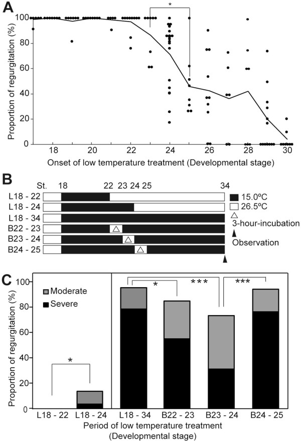
Figure 3.
Proportion of blood regurgitation phenotype after incubation at 15°C during embryogenesis. A: Proportion of embryos showing regurgitation at st.34 after the onset of the 15°C treatment at the stages indicated (st.17, n = 50; st.18, n = 50; st.19, n = 54; st.20, n = 69; st.21, n = 53; st.22, n = 80; st.23, n = 66; st.24, n = 79; st.25, n = 65; st.26, n = 98; st.27, n = 50; st.28, n = 62; st.29, n = 86; st.30, n = 62). Only clutches with fertilized eggs (n ≥ 4) were used for the assay. Dots indicate each clutch. Lines indicate the average of proportions in clutches at the same stage. B: Scheme of the 15°C -treatment experiment used in C. Solid bar, 15°C; open bar, 26°C. L: Low-temperature treatment, B: Breakage of 15°C treatment. C: Comparison of the proportion of regurgitation between embryos at various stages (L18–22 (n = 189), L18–24 (n = 208), B22–23 (n = 40), B23–24 (n = 64), and B24–25 (n = 68)). Gray columns, moderate regurgitation; black columns, severe regurgitation. The classification of the regurgitation phenotype was performed by visual inspection. Asterisks indicated the level of significance (*: p < 0.05; ***: p < 0.005).

