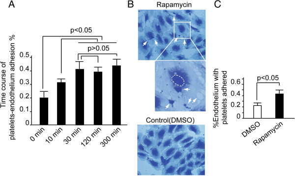
Figure 2.
Rapamycin induced platelets-endothelial adhesion. HUVECs were cultured on the gelatin-coated coverslips. Freshly isolated platelets were added to the coverslip and incubated for the indicated time. Cells were fixed and stained with crystal violet. The coverslips were mounted and imaged by microscopy. (A) The time course of platelet-endothelial adhesion exhibited that the platelets-endothelial adhesion reached its plateau at the 30th minute. Columns, means of three independent experiments; bars, SD. (B) The upper and middle panel indicated that platelets were seen adhering on the HUVECs. (C) HUVECs with platelets adhered were calculated and significant difference existed between the two groups (P < 0.05). Columns, means of three independent experiments; bars, SD.

