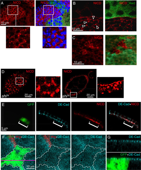
Figure 5.
Defective intracellular distribution of Notch in awd mutant cells. (A-B) Stage 8 egg chambers were dissected from hs-flp; +/+; Ubi-GFP, FRT82B/FRT82B, awdj2A4 females, and stained for NICD (red), Awd (green) and DNA (blue). Notch over-accumulates in vesicles near the cell periphery (insets in (A) and arrowheads in (B)). (C) The wing disc was dissected from hs-flp; +/+; Ubi-GFP, FRT82B/FRT82B, awdj2A4 third instar larva and stained for NICD (red). awd clones were identified by lack of Awd staining (pseudo-colored in green). Notch in awd mutant clones accumulates in large vesicles. (D) Surface and cross-section views of shits stage 7 egg chambers from females incubated at 29°C and stained for NICD. Very large aggregates are seen on the surface and throughout the cells. (E) A stage 7 egg chamber from a hs-flp/GbeSu(H)m8-lacZ; act-Gal4, UAS-GFP/+; FRT82B, act-Gal80/FRT82B, awdj2A4 female was stained for DE-cadherin (cyan) and NICD (red). Notch accumulates in awd mutant cells (GFP-positive) that show normal DE-cadherin distribution. (F-G) Third instar wing imaginal disc dissected from a hs-flp/+; act-Gal4, UAS-GFP/+; FRT82B, act-Gal80/FRT82B, awdj2A4 larva and stained for DE-cadherin (cyan) and Wg (red) in which the awd mutant clone is marked by GFP expression and outlined in F by the dotted area. (F) The confocal section of the apical region of disc cells (x-y) shows that awd loss of function does not affect the distribution of DE-cadherin. (G) The cross section through the disc epithelium (x-z) with apical side up also shows an unaffected apical/basal polarity distribution of DE-cadherin in awd mutant cells. The pink line indicates the position of the x-z section. Awd, Abnormal wing discs; NICD, Notch intracellular domain.

