Figure 1.
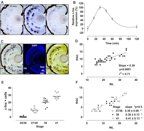
Light induces c-fos expression in the inner nuclear layer (INL) and retinal ganglion cell (RGC) layer as early as Stage 37/38. (A)c-fos in situ hybridization of transverse sections from Stage 42 tadpole eyes that developed in the dark and were exposed to light (2500 lux) for the indicated times. (B) Graph of the integral optical density of c-fos in the eye (mean ± SEM; n = 8 eyes) relative to that measured after 0 (0%) or 30 (100%) minutes of light exposure. (C)c-fos in situ hybridization (left), DAPI staining (middle), and corresponding merged picture (right) of a representative central section used to quantify c-fos + cells in the INL and the RGC layer. (D) Correlation between the numbers of c-fos-expressing cells in the INL and the RGC layer. Data for each central retina quantified are represented by a dot (n = 33). The linear regression and the coefficient of regression are indicated. (E) Embryos at different stages of development were exposed to light (2500 lux, 30 minutes) and the number of c-fos + cells in a section from the central retina quantified. Differences between all stages analyzed were statistically significant (P < 0.05; one-way ANOVA, Bonferroni’s multiple comparisons test). Line indicates the mean. (F) Correlation between the numbers of c-fos + cells in the INL and the RGC layer. The slope for each group is indicated (P value for each linear regression from Stages 35/36 and older are statistically significant; P < 0.05; there were no significant differences in the slopes between different stages). ONL, Outer nuclear layer; ON, Optic nerve. Scale bar = 50 μm.

