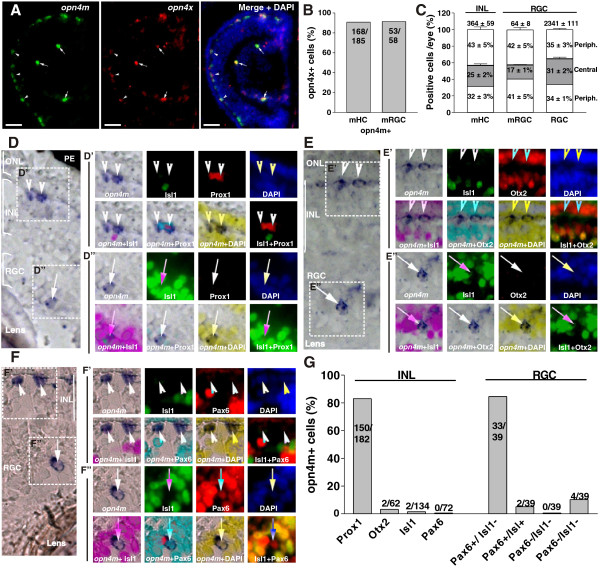
Figure 3.
Expression of melanopsin (opn4m and opn4x) in the Xenopus tadpole retina. (A) Double in situ hybridization against opn4m and opn4x in a transverse section of the retina of a Stage 42 tadpole. Merged photograph with DAPI staining is shown (right). Double-positive cells in the outer nuclear layer (ONL) (melanopsin-expressing horizontal cells (mHCs); arrowheads) and in the retinal ganglion cell (RGC) layer (melanopsin-expressing retinal ganglion cells (mRGCs); arrows) are indicated. Scale bar = 100 μm. (B) Quantification of the number of opn4x + cells that also expressed opn4m, shown as percentage. The number of cells counted is indicated. (C) Distribution of mHCs and mRGCs counted in consecutive sections throughout the whole eye, divided into two peripheral domains and one central domain. The percentage of cells located in each domain, and the total numbers of cells counted (mean ± SD; n = 3 eyes), are indicated. The total number of RGCs counted (based on DAPI + nuclei), and their distribution, is also shown. (D-F)In situ hybridization identified opn4m + cells in the outer segment of the INL (D′-F′, arrowheads) and in the RGC layer (D′-F′, arrows). In situ hybridization was followed by immunohistochemistry against Isl1 (green) or Prox1 (red) (D′ and D′′), Isl1 and Otx2 (red) (E′ and E′′), or Isl1 and Pax6 (red) (F′ and F′′). Nuclei stained with DAPI (blue) and merged photograph of the corresponding images are presented. (G) The percentage of cells double-labeled for opn4m and the indicated marker in the INL and the RGC layer, as well as the number of cells counted, are shown.

