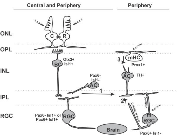
Figure 7.
Neuronal circuit diagram of the light input pathway in the tadpole retina. Cells expressing c-fos in response to a first exposure to light are shown in gray, and we propose that they correspond to second-order or third-order neurons. Photosensitive cells (>>>) do not express c-fos as they are first-order neurons. Melanopsin-expressing retinal ganglion cells (mRGCs) induce c-fos in response to light, either by serving as first-order neurons because of their intrinsic photosensitivity, or via a role as a second-order or third-order neuron that receives synaptic inputs from other retinal cells. The classic IF circuit is present in both the central and peripheral retina. In lower vertebrates, rods (R) and cones (C) in the ONL are connected via gap junctions (black ovals) and synapse directly on a single class of On-BCs (Otx2+ / Isl1+), which drive activity in a sub-population of ACs (Pax6− / Isl1−) [59]. Finally, the pathway drives c-fos expression in two equally abundant RGC sub-populations (Pax6− / Isl1+ and Pax6+ / Isl1+). Not illustrated are the cells of the retinal IF circuit that do not express c-fos in response to light: the HCs, Off-BCs, and Pax6+ and/or Isl1+ ACs. The cells involved in non-image-forming (NIF) tasks express melanopsin and are preferentially distributed in the peripheral retina. These include the mHCs (Prox1+),mRGCs (Pax6+ / Isl1−), and dopaminergic (TH+) ACs that turn on c-fos with blue light. Three possible connections may induce c-fos expression in the TH + ACs: 1) PR-initiated inputs from On-BCs to mRGCs and/or TH + ACs; 2) synaptic interaction between an mRGC axon collateral with INL cells [62] to provide a retrograde signal from mRGCs to TH + ACs [63]; and 3) a circuit that may only exist in lower vertebrates, whereby mHCs act as first-order neurons, and interplexiform (TH+) ACs link mHCs to mRGCs.

