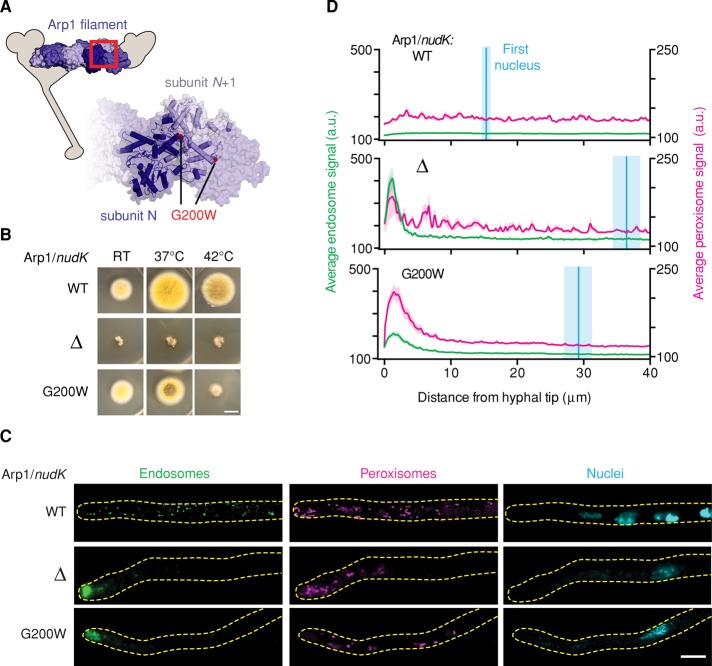
FIGURE 2:
The Arp1 allele is temperature sensitive. (A) Structural model of the Arp1 minifilament based on the structure of Oryctolagus cuniculus skeletal muscle F-actin (PDB 3G37; Murakami et al., 2010), illustrating the predicted position of the G200W mutation in Arp1. Inset, enlargement of the region indicated by the red box. (B) Images of colony morphology show that the Arp1G200W mutation is temperature sensitive. Wild-type, Arp1Δ, and Arp1G200W strains were grown at room temperature, 37°C, or 42°C. The wild-type strain forms large colonies that conidiate at all temperatures (top). The Arp1Δ strain forms small colonies that do not conidiate at any temperature tested (middle). The Arp1G200W mutant conidiates at room temperature and shows a minor conidiation defect at 37°C, but the colonies are small and nonconidiating at 42°C (bottom). Scale bar, 1 cm. (C) Representative micrographs of strains grown at 42°C. In wild-type hyphae, endosomes, peroxisomes, and nuclei were distributed (top). In both Arp1Δ and Arp1G200W hyphae, endosomes and peroxisomes accumulated at the hyphal tip, and the distance between the distal nucleus and hyphal tip was increased (middle and bottom, respectively). Scale bar, 5 μm. (D) Endosome (green) and peroxisome (magenta) distribution was quantified using line scans of fluorescence micrographs. Mean (green or magenta lines) ± SEM (green or magenta shading) fluorescence intensity as a function of distance from the hyphal tip. Arp1Δ and Arp1G200W hyphae showed a pronounced peak of fluorescence intensity for endosomes and peroxisomes in the hyphal tip, whereas wild-type cells did not. Nuclear distribution was quantified by measuring the distance from the distal nucleus to the hyphal tip. The mean (cyan line) ± SEM (cyan shading) distance was 15.30 ± 0.79 μm (n = 19) in wild-type, 36.48 ± 5.74 μm (n = 24) in Arp1Δ, and 29.23 ± 2.19 μm (n = 32) in Arp1G200W hyphae. The nuclear distributions in both Arp1Δ and Arp1G200W hyphae were significantly different from that in wild-type hyphae (p < 0.001, unpaired t test).

