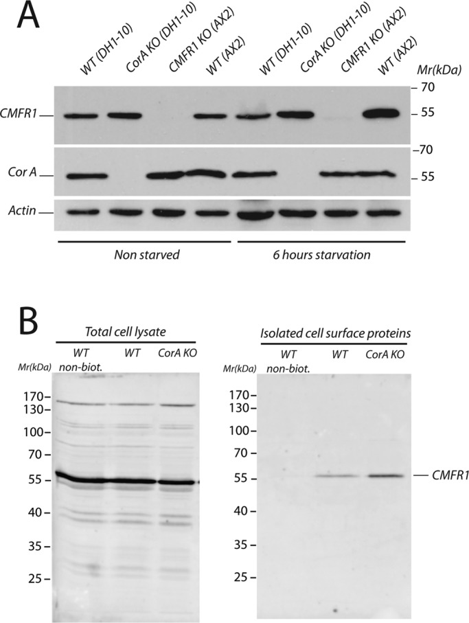
FIGURE 7:

CMF receptor 1 expression in wild-type and coronin A–deficient cells. (A) Exponentially growing wild-type (DH1-10 and AX2), coronin A–deficient, and CMFR1-deficient cells were washed in BSS, and membranes were extracted immediately or after 6 h of starvation in BSS at a density of 10 × 106 cells/ml. Equal amounts of cells were separated by 10% SDS–PAGE and analyzed by Western blot for CMFR1, coronin A, and actin expression. (B) Wild-type and coronin A–deficient cells were cooled, pelleted, and washed twice. Cell surface proteins were then biotinylated and purified. A Western blot against CMFR1 was performed on total cell lysates (left) or isolated cell surface proteins (right).
