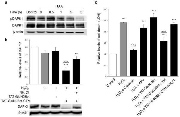
Figure 5. TAT-GluN2Bct-CTM knocks down H2O2-activated DAPK1, protecting neurons against H2O2-induced neurotoxicity in neuronal cultures.
(a) Immunoblotting for phosphorylated DAPK1 (pDAPK1) revealed a time-dependent activation (dephosphorylation) of DAPK1 by H2O2 treatment (300μM; 30min; n=4). (b) Bath application of 100μM TAT-GluN2Bct-CTM (36.54±7.1% of control; n=8; p=0.001), but not TAT-GluN2Bct (89.13±10.78%; n=7; p=0.311), 60min prior to and during H2O2 treatment (300μM; 30min) knocked down DAPK1 at 2hrs post-washout, which was rescued by NH4Cl (20mM; n=8; p=0.003 to control*; p=0.106 to H2O2-treatedΔ). One-way ANOVA, F(4,34)=11.628, p<0.001. Bars represent DAPK1 levels relative to naïve group. β-actin was used as a loading control. (c) LDH assay revealed that H2O2 treatment (300μM; 30min) resulted in a significant increase in neuronal death 12hrs after treatment (n=8; 2.50±0.12; p<0.001 to control), which was rescued by breaking down H2O2 with catalase (100U; n=4; 1.17±0.02; p=0.001 to H2O2 group). H2O2-induced neurotoxicity was significantly reduced by TAT-GluN2Bct-CTM (50μM; applied 60min prior to and maintained throughout the experiments; n=9; 1.56±0.08; p=0.001 to H2O2 group), but not by TAT-GluN2Bct (50μM; n=9; 2.63±0.10; p=0.105 to H2O2 group) or the NMDAR antagonist APV (1μM; n=4; 2.16±0.14; p=0.169 to H2O2 group). NH4Cl abolished the neuroprotective effect of TAT-GluN2Bct-CTM (n=4; 2.39±0.27; p=0.538 compared to H2O2 group). One-way ANOVA, p<0.001, F(6,41)=26.842. *,Δ p<0.05, **,ΔΔ p<0.01 and ***, ΔΔΔp<0.001; bars represent relative mean values±s.e.m. normalized to the naïve control (white bar, arbitrarily set as 1). n represents individual experiments from at least 3 separate primary cultures. Full-length blots are presented in Supplementary Figure 9.

