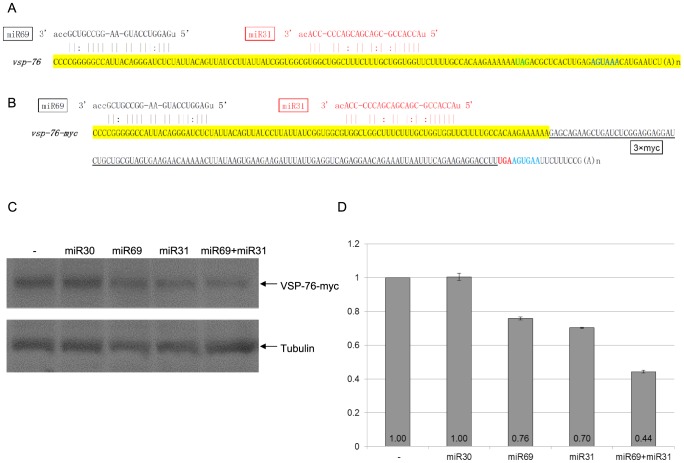
Figure 4. miRNA-mediated regulation of VSP-76 expression.
A) Schematic diagram of the vsp-2 mRNA 3′ region (100 nts upstream of the stop codon and the 3′ UTR) with the binding sites for miR24 and miR56. Coloring is as in Figure 3A. B) Schematic diagram of the vsp-76-myc mRNA detailing incorporation of the 3xmyc tag and elimination of the native 3′ UTR. C) Giardia cells expressing vsp-76-myc transfected with the indicated miRNAs was analyzed with Western blot. Tubulin was used as a loading control. D) Densitometry analysis of three independent Western blot experiments is shown as a histograph with the error bars representing the standard deviation. Expression of VSP-76-Myc is repressed in the presence of miR69 or miR31 to 76% and 70% of the control, respectively. The presence of both miRNAs represses VSP-76-Myc expression down to 44%, indicating cooperative miRNA action. miR30, which does not target vsp-76, does not affect VSP-76-myc expression.

