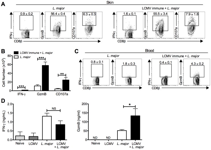
Figure 4. CD8 T cells infiltrating the leishmanial lesions express gzmB but low levels of IFN-γ.
B6 mice were infected with LCMV Armstrong or left uninfected. After 30 days, mice were infected with L. major. After 4 weeks, infected ears (A) and peripheral blood (C) were taken for analysis by flow cytometry. Cells from the infected ears were incubated with BFA, monensin and CD107a antibody for 5 hours and then stained for additional cell surface markers and intracellular proteins. Representative dot plots (A) and total cell numbers (B) are shown. Peripheral blood was taken and white blood cells were isolated and stained for cell surface markers and intracellular proteins. Representative dot plots are shown (C). Whole ear tissue was homogenized and supernatants were analyzed for gzmB and IFN-γ by ELISA (D). Flow data are representative of five independent experiments (n = 4–5 mice per group). Ear supernatant data are representative of two independent experiments (n = 4 mice per group). Percentages are shown as mean ± SEM.

