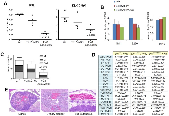
Figure 2. Disruption of hematopoiesis in Evi1δex3/δex3 newborn mice.
(A,B) Flow cytometric profiles of wild type, Evi1δex3/+ and Evi1δex3/δex3 littermate fetal livers at E14.5. (A) HSC and progenitor cell subpopulations were detected by a combination of markers (KSL: c-Kit+, S: Sca-1+, L: lineage−, or KL-CD34+). We found a significant reduction of cells in the Evi1-deleted samples; p values are from an unpaired t-test between +/+ and Evi1δex3/δex3 fetal livers. (B) Bar graph shows the number of granulocytes (Gr1), B-lymphocytes (B220) and erythroid cells (Tert119) in fetal livers of various different genotypes. (C) Colony forming counts from cells of 3 fetal livers of each genotype at E14.5 We observed a significant reduction in colony formation between +/+ and Evi1δex3/δex3 fetal livers, p = 0.0057 (unpaired t-test). No BFU-E or CFU-Mix colonies were identified. (D) Hemogram results for 4 hr- to 24 hr-old wild type (N = 17), Evi1δex3/+, (N = 30) and Evi1δex3/δex3 (N = 16) littermate pups. Mean ± SEM is indicated. *p<0.05, **p<0.01, ***p<0.001, unpaired t-test. Leukocyte counts in peripheral blood and white blood cell differentials reveal a mild leucopenia in Evi1δex3/δex3 newborn mice. Platelet (PLT) counts and mean platelet volume (MPV) results show a mild hypoproliferative thrombocytopenia in Evi1δex3/δex3 pups. Normal erythrocyte counts, hemoglobin quantification and hematocrit assessment in the peripheral blood of Evi1δex3/δex3 animals. Mean corpuscular volume (MCV), mean corpuscular hemoglobin (MCH), mean corpuscular hemoglobin concentration (MCHC) and red cell distribution widths (RDW) are shown. (E) Hematoxylin and eosin staining of 5 µm sections of 24 hr- to 48 hr-old Evi1δex3/δex3 pups. Mild hemorrhages were seen in 31% of the mice (4 out of 13 pups).

