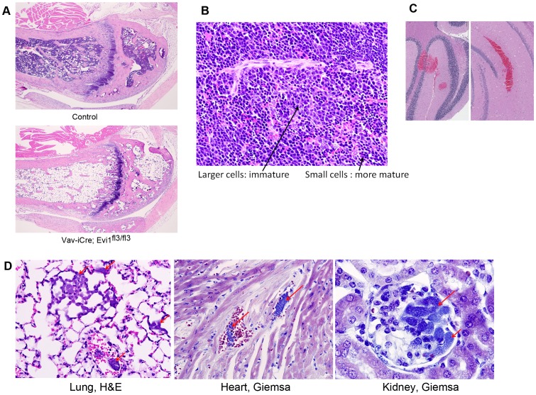
Figure 4. Spontaneous lethal bone marrow depletion in mice harboring an Evi1 exon3 deletion in the hematopoietic system.
(A) Histology was performed on sick Vav-iCre; Evi1fl3/fl3 and littermate control mice. Bone marrow depletion was observed in the mutant mice. Adipose tissue replaced the hematopoietic cells in the bone marrow. (B) Increased erythropoiesis in the spleen of Vav-iCre; Evi1fl3/fl3 mice. No visible border was found between the red pulp and white pulp. Erythroid cells are shown by the arrows. Excess erythropoiesis in spleen likely happens to compensate for bone marrow loss. (C) H&E stained sections of the brain of a dying Vav-iCre; Evi1fl3/fl3 mouse. Hemorrhages (red areas) were visible at several locations (also see Fig. S3E in File S1). (D) Histological sections of tissues from dying Vav-iCre; Evi1fl3/fl3 animals showing bacteremia. Red arrows indicate the presence of bacteria in alveolar capillaries. Giemsa stains reveal the presence of cocci or small rods within glomerular capillaries. No sign of immune system defense (inflammatory cells) was observed despite the infection.

