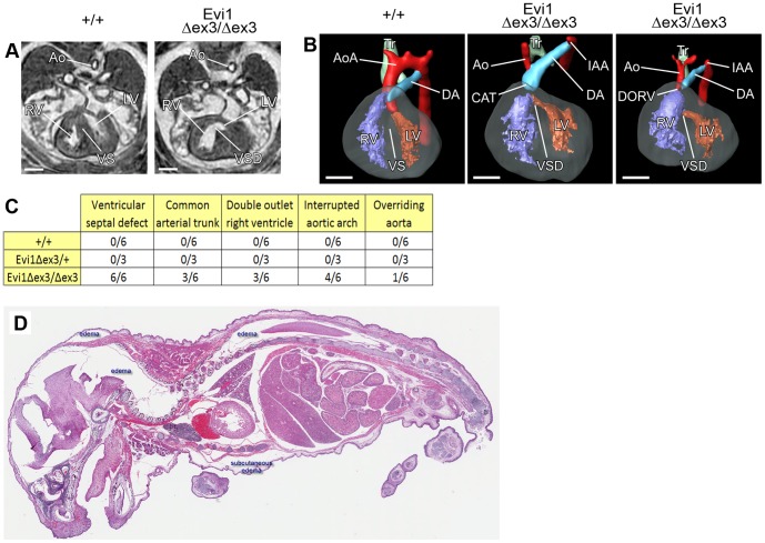
Figure 5. Cardiac malformations and failure in Evi1δex3/δex3 mice.
(A) Transverse sections and (B) 3D reconstruction (left-ventral oblique view) of hearts from Evi1δex3/δex3 or wild type littermate (+/+) E15.5 embryos analyzed by magnetic resonance imaging (MRI). The aorta (Ao), right ventricle (RV), left ventricle (LV), ventricular septum (VS), trachea (Tr), aortic arch (AoA) and ductus arteriosus (DA) are indicated. Ventricular septal defect (VSD), interrupted aortic arch (IAA) and common arterial trunk (CAT) were observed in Evi1δex3/δex3 hearts. (C) List of the congenital heart defects identified in fifteen E15.5 embryos of various different genotypes by MRI and 3D reconstruction. (D) Hematoxylin and eosin staining of 5 µm sections of a sick Evi1δex3/δex3 pup. Subcutaneous and other tissue edema (white spaces) was present, consistent with heart failure.

