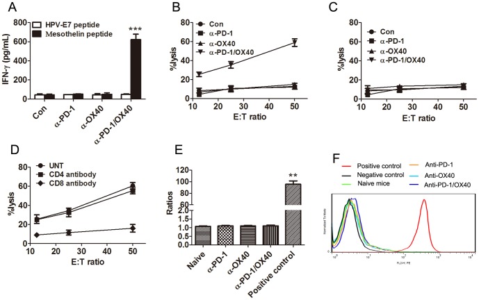
Figure 5. Mice treated with anti-PD-1/OX40 mAbs developed a tumor antigen-specific CTL response.
A, Mice injected i.p. with 1×10⁶ ID8 cells 10 day earlier were injected thrice at 4 days interval with 200 µg of control, single or combined anti-PD-1/OX40 mAb. Seven days after the last mAb injection, splenocytes from treated mice were cultured in the presence or absence of H-2Db-restricted mesothelin-derived peptides or control HPV-E7-derived peptide for 3 days and IFN-γ production in the supernatants were assayed by ELISA. B, Pooled splenocytes (5×10⁶) from all mice treated with control, single or combined anti-PD-1/OX40 mAb were incubated with 5×10⁵ UV-irradiated ID8 cells for 4 days prior to subject to analysis of antigen-specific CTL activity by CytoTox 96 Non-radioactive cytotoxicity assay using EL4 cells pulsed with H-2Db-restricted mesothelin peptide as target cells. C, As a specific control, pooled splenocytes were tested cytotoxicity against EL4 target cells pulsed with control HPV-E7 peptide. D, The killing activity of splenocytes from combined mAb treated mice on mesothelin-pulsed EL4 cells was also evaluated in the presence of blocking anti-CD4, anti-CD8 or control antibody. Data were expressed as M±SEM of triplicate wells in A-D. E, Sera were obtained from naïve mice (5 mice), single mAb treated mice (5 mice each group) or 2 mAb treated long-term surviving mice (4 mice). The presence of mesothelin-specific antibodies was determined by flow cytometry via staining mesothelin-expressing ID8 cells with collected sera in a 1∶200 dilution, followed by staining with secondary PE-conjugated anti-mouse IgG antibody and MFI was calculated. Staining with commercially available anti-mesothelin antibody plus secondary antibody or secondary antibody alone was used as positive or negative control. The ratios of MFI from positive control or sera to MFI from negative control were shown. F, A representative histogram in E was shown.

