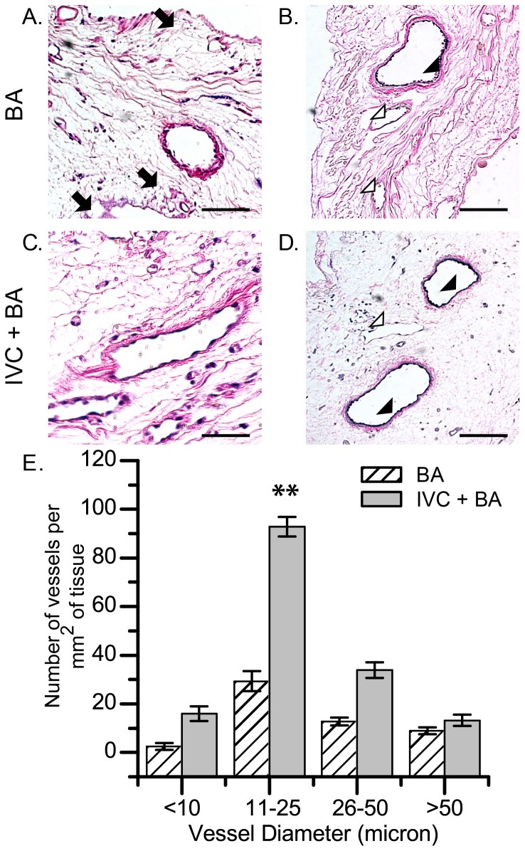
Figure 2. Histologic assessment of decellularized rat heart scaffolds seeded with RAECs.
(A,C) H&E and (B,D) Verhoeff-Van Gieson staining of scaffolds recellularized by the BA only technique (A,B) and the combined IVC+BA technique (C,D); arrows indicate cell-free vessels (A), arrow heads indicate elastin-positive vessels with cell nuclei (B,D), and open arrow heads indicate elastin-negative vessels (B,D). All scaffolds were recellularized with 4×10⁷ RAECs. (E) Quantification of the number of vessels lined by DAPI-positive cell nuclei in the mid-ventricular wall; the results are grouped according to vessel diameter (n = 3 per data set; mean ±SEM). **p<0.001 for IVC+BA vs BA re-endothelialization techniques for vessels with a diameter of 11–25 microns. Scale bars represent 125 microns (A–D).

