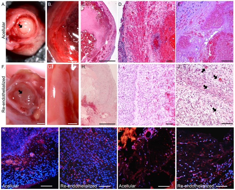
Figure 5. Characterization of heterotopic transplants.
(A–E) Acellular and (F–J) re-endothelialized scaffolds seven days after heterotopic transplantation. Short axial view of an aorta with blood clot (arrow) from an acellular scaffold (A) and a non-clotted aorta (arrow) from a re-endothelialized scaffold (F) after transplantation. Short axial view of the ventricle wall of an acellular scaffold with a blood clot (B) and a re-endothelialized scaffold (G). H&E staining of a transplanted acellular scaffold (C–E) with a blood clot inside the ventricular cavity and a re-endothelialized scaffold (H–J) at increasing magnification (2X, 10X, and 20X). Arrows point to patent vessels with and without blood (J). CD31 (K) and VEGFR2 (L) (red) staining in transplanted scaffolds; DAPI-positive nuclei are blue. Scale bars represent 1 mm (A–C and F–H) and 100 microns (D, E, I, J, K, and L).

