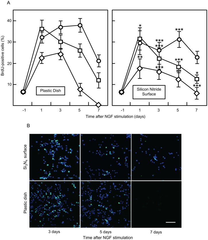
Figure 9. Assessment of PC12 cell proliferation using a BrdU assay.
(A) Cells were seeded on different surfaces and 24 hours after seeding, they were stimulated with NGF (50 ng/ml). After 2 hours incubation in growth medium containing BrdU solution, cells were fixed, permeabilized and stained using an Alexa Fluor 488 conjugated anti-BrdU antibody. Finally, BrdU positive cells were counted in 20 samples and the proportion (%) of labeled cells per counted field was calculated. Standard error is shown using error bars. The following media were tested: FBS (+) NGF (−) (○); FBS (+) NGF (+) (□); and FBS (−) NGF (+) (⋄). (*, p<0.05; * *, p<0.01; * * *, p<0.001 vs. plastic culture dish). (B) Cells were seeded on different surfaces and cultured in the absence of serum and with NGF treatment. They were fixed using a paraformaldehyde-based solution and immunocytochemical detection of the BrdU antigen was performed to identify proliferative (S phase) cells. Images were captured at different time points. Representative images of each condition are shown. Scale bar: 50 µm.

