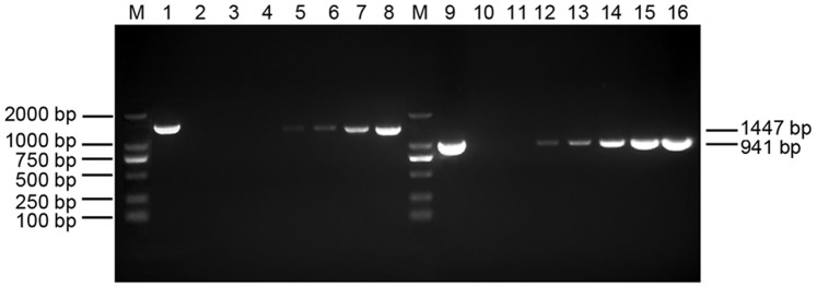
Figure 1. Detection of WSSV in hemocytes of F. chinensis post WSSV infection.

Lane M, marker; Lane 1–2, the first-step PCR products of positive control and negative control; Lane 3–8, the first-step PCR products of hemocytes at 0, 6, 12, 24, 36 and 48 hpi; Lane 9–10, the second-step PCR products of positive control and negative control; Lane 11–16, the second-step PCR products of hemocytes at 0, 6, 12, 24, 36 and 48 hpi.
