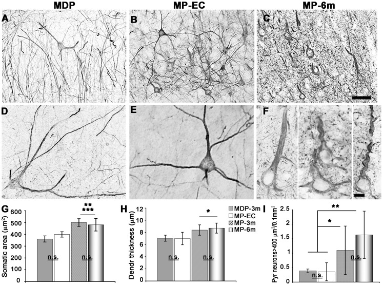
Figure 3. Dysmorphic neocortical pyramidal neurons in epileptic MP rats.
A–F) Low- (A–C) and high-power (D–F) microphotographs of NF200+ enlarged pyramidal neurons in MDP (A, D) and epileptic MP-EC (B, E) and MP-6m (C, F) rats. Note the progressive perikaryal enlargement and dendritic simplification of dysplastic neurons in MP-6m rats vs both non-epileptic MDP and early-chronic epileptic MAM rats. G–I) Quantification and statistical analysis of somatic area (G), dendritic thickness (H) and cell numbers (I) of NF200+ enlarged pyramidal neurons (≥400 µm2) at different epilepsy stages in MAM rats (*p<0.05; **p<0.01; ***p<0.001; n.s: not significant). For somatic area and dendrite thickness at least 4 animals/each group were analyzed (25 neurons per animal). Scale bars: 100 µm in A–C; 10 µm in D–F.

