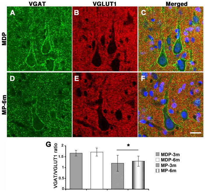
Figure 6. Altered synaptic input on neocortical pyramidal neurons of epileptic MP rats.
A–F) VGAT+ (green) and VGLUT1+ (red) synaptic terminals on pyramidal neurons from non-epileptic MDP (A–C) and epileptic MP-6m rats (D–F). Panels C and F are merged images counterstained with Neurotrace™ (blue) to reveal neuronal nuclei. G) Quantification of VGAT+ and VGLUT1+ peri-somatic and -dendritic terminals on neocortical pyramidal neurons expressed as VGAT/VGLUT1 IF ratio. VGAT/VGLUT1 ratio was significantly decreased in MP vs aged-matched MDP control (*p<0.05 MP-3m/6m vs MDP-3m/6m). No differences were found in MDP-3m vs MDP-6m and MP-3m vs MP-6m. Scale bar: 20 µm.

