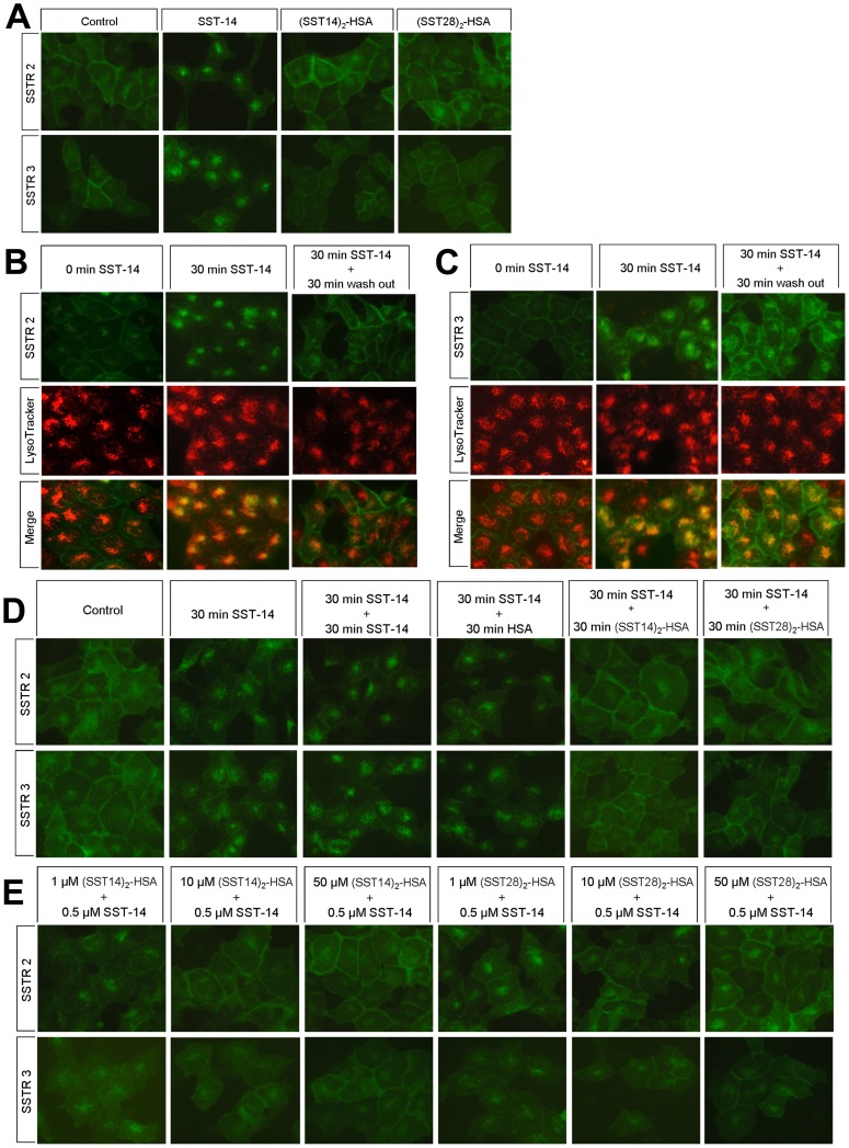
Figure 3. Regulation of somatostatin-HSA fusion proteins to receptors internalization and recycling in SSTR2- and SSTR3-expressing cells.
(A) Comparison of agonist-induced endocytosis of SSTR2 and SSTR3. HEK 293 cells stably expressing SSTR2- or SSTR3-EGFP were exposed to 1 µM SST-14, (SST14)2-HSA or (SST28)2-HSA for 0 or 30 minutes. The subcellular distribution of receptor protein was examined by fluorescence microscopy. (B, C) Internalization and recycling of SSTR2 and SSTR3. HEK 293 cells expressing SSTR2- or SSTR3-EGFP were exposed to 0.5 µM SST-14 for 30 minutes. Cells were then washed and followed by an agonist-free interval of 30 minutes. Cells were stained with LysoTracker Red DND-99 according to the manufacturers’ protocols. Locations of SSTR2 or SSTR3 (upper panel, green) and lysosomes (middle panel, red) were examined by fluorescence microscopy. The merged image (lower panel) shows co-localization (yellow) of SSTR2 or SSTR3 and lysosomes. Images were processed using ImageJ software. (D) Fusion proteins accelerated the recycling of internalized receptors to the plasma membrane. SSTR2- or SSTR3-EGFP-expressing HEK 293 cells were treated by 0.5 µM SST-14 for 30 minutes, then 50 µM of SST-14, HSA, (SST14)2-HSA or (SST28)2-HSA were added with little change in final volume. After 30 minutes treatment, the subcellular distribution of receptor protein was examined by fluorescence microscope. (E) Dose-dependent inhibition of receptors internalization. SSTR2- or SSTR3-EGFP-expressing HEK 293 cells were exposed to 1 µM, 10 µM or 50 µM (SST14)2-HSA or (SST28)2-HSA for 30 minutes, then 0.5 µM SST-14 were added while final volume changed little. After 30 minutes treatment, the subcellular distribution of receptor was examined by fluorescence microscope. Representative results from one of three independent experiments performed in duplicate are shown.

