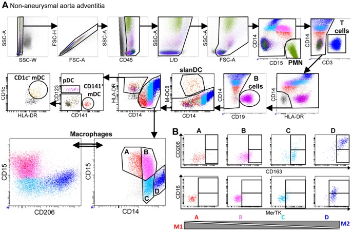
Figure 2. Analysis of leukocyte subsets in the adventitia from a non-aneurysmal abdominal aorta by polychromatic flow cytometry.
(A) The gating strategy to analyze CD45+ leukocytes from a non-aneurysmal abdominal aorta by 15-color flow cytometry is shown. This strategy allows the simultaneous definition of CD14−CD15hi polymorphonuclear cells (PMN), CD3+ T cells, CD19+ B cells, CD14+ macrophages, and dendritic cell DC) subsets comprising CD14loM-DC8+ slanDC, and among lin−HLA-DR+ events, CD123+ plasmacytoid DC (pDC), and two subsets of myeloid DC (mDC), CD141+ mDC and CD1c+ mDC. The lower right dot plot shows four macrophage subsets defined by their relative expression of CD14 and CD15, and the lower left panel shows CD206 and CD15 expression by these macrophage subsets. (B) Flow cytometric dot plots of CD163 and CD206 (upper dot plots) and MerTK and CD16 (lower dot plots) expression by the four macrophage subsets defined in (A).

