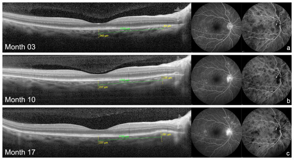
Figure 1.

Choroidal bulging and angiographic findings in the RE of case 2. Absence of abnormalities on EDI-OCT at 3 months of disease in RE (a). Gradual increase in choroidal thickness (choroidal bulging) in the outer nasal macular sub-field at months 10 (b) and 17 (c) was associated with gradual increment in optic disc hyperfluorescence on FA and localized appearance of dark dots on ICGA (arrows). Fluctuation in the diffuse density of dark dots on ICGA was concomitantly observed.
