
FIGURE 5.
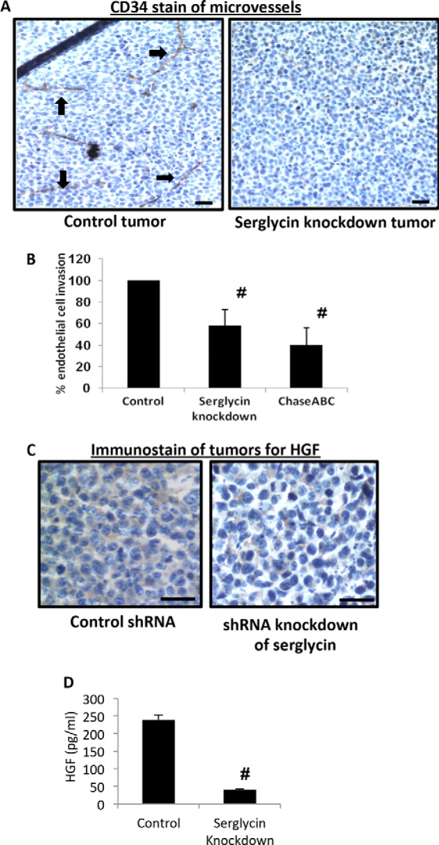
Tumors formed by serglycin-knockdown cells have a poorly developed vasculature and decreased levels of human HGF. A, immunohistochemistry of tumors formed by control (left panel) or serglycin-knockdown (right panel) cells. Tumor tissue was stained with antibody to CD34 (brown) and counterstained with hematoxylin (blue). Microvessels are indicated with black arrows. Scale bars, 200 μm. B, conditioned medium from serglycin-knockdown cells inhibits endothelial invasion. Endothelial cells were placed in the upper chamber of Matrigel invasion chambers and conditioned medium from control or serglycin-knockdown cells or from control cells pretreated with Chase ABC was placed in the lower chamber. After overnight incubation, endothelial cells that invaded through the Matrigel were fixed, stained, and counted. Data are mean ± S.D. of three independent experiments. #, p < 0.05 compared with control. C, immunohistochemistry of sections from xenograft tumors formed from control (left panel) or serglycin-knockdown (right panel) cells stained for human HGF (brown) and counterstained with hematoxylin (blue). Scale bars, 200 μm. D, medium conditioned by myeloma cells for 48 h was collected, and levels of human HGF quantified by ELISA. Data are from mean ± S.D. from three separate experiments. #, p < 0.05 compared with control.