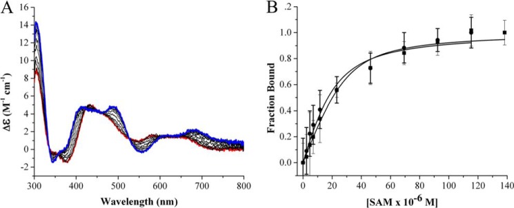
FIGURE 2.

A, visible region circular dichroism of PFL-AE showing the [4Fe-4S] cluster is perturbed upon AdoMet binding under anaerobic conditions. Red, as isolated PFL-AE; blue: PFL-AE with AdoMet (SAM) fully bound. The PFL-AE concentration was 50 μm, and AdoMet concentrations were in the range of 0–140 μm; the assay was performed in 20 mm HEPES, 250 mm NaCl, 1 mm DTT, pH 7.4 at 25 °C. B, AdoMet binding data for PFL-AE (●) and AdoMet binding to the PFL-AE/PFL complex (■). PFL-AE and PFL (when present) concentrations were 50 μm each; other buffer conditions were as indicated for A. The data were analyzed using changes in ellipticity at 400 nm divided by total change in ellipticity, which was then plotted as function of AdoMet concentration and fit to the quadratic binding equation. CD parameters were set to a sensitivity of 100 millidegrees, with a data pitch of 0.1 nm, in continuous scan mode with a scan rate of 100 nm/minute, a scan range of 300–800 nm, response of 1 s, bandwidth of 1.0 nm, and an accumulation of three scans; all measurements were performed using a 1-cm path length anaerobic cuvette.
