FIGURE 2.
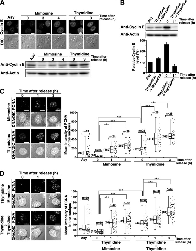
Mimosine-induced synchronization at the G1/S phase boundary. HeLa S3 cells were synchronized by the indicated methods and released for the indicated times. A, cells were fixed in methanol and stained for cyclin E. DIC, differential interference contrast. Bottom panel, cell lysates were analyzed by Western blotting. Asy, asynchronous cells. Scale bars = 10 μm. B, cell lysates were analyzed by Western blotting. G1 phase cells were prepared by 14-h release from Thy → Thy treatment. The results (percent) represent the mean ± S.D. from three independent experiments (bottom panel). C and D, the Triton-resistant fraction was fixed and stained for PCNA and DNA. Right panels, mean fluorescence intensities of PCNA immunostaining in the nucleus were plotted. ***, p < 0.001, calculated by Student's t test.

