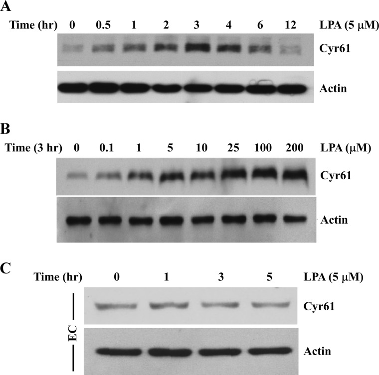
FIGURE 1.
LPA markedly induced Cyr61 protein expression in SMCs. A, time course of LPA induction of Cyr61 protein expression in mouse aortic SMCs. Cultured cells were starved for 48 h prior to LPA (5 μm) stimulation for various times as indicated. Cell lysates were subjected to Western blot analysis. The same membrane was reprobed with β-actin antibody to assess protein loading. B, Western blot analysis showing dose-dependent LPA induction of Cyr61 expression in SMCs. C, Western blot analysis showing the effect of LPA on Cyr61 expression in human aortic endothelial cells (EC). β-Actin served as the loading control.

