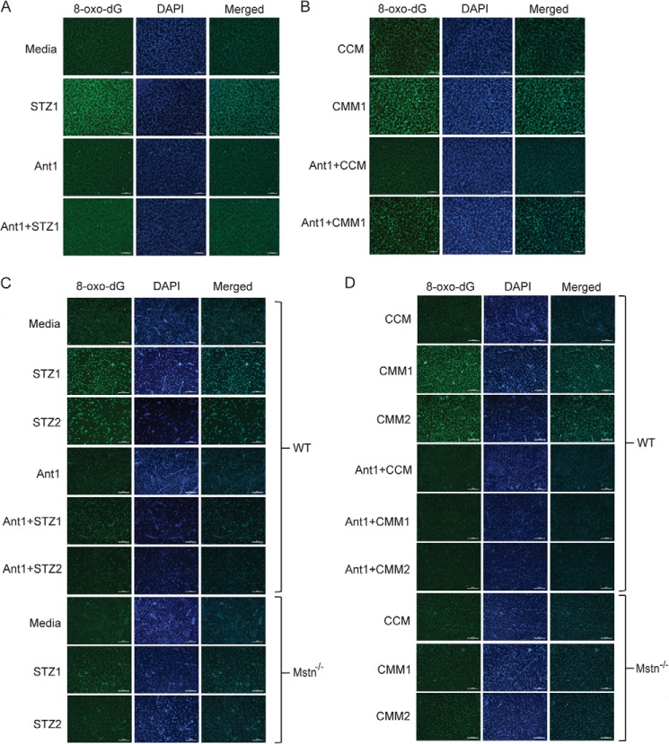
FIGURE 5.
STZ-induced DNA damage in proliferating myoblasts is attenuated by antagonizing Mstn. A, immunocytochemistry was performed for 8-oxo-dG on proliferating C2C12 myoblasts treated for 48 h with or without STZ1 and with or without pre-treatment of Ant1. The fluorescence for 8-oxo-dG (green) and DAPI (blue) was viewed under a Leica upright microscope and images were taken at 10× magnification. Increased fluorescence is directly proportional to increased DNA damage; scale bar represents 100 μm (n = 3). B, 8-oxo-dG immunostaining in proliferating C2C12 myoblasts treated for 48 h with CCM, CMM1, Ant1+CCM or Ant1+CMM1. Images were taken as mentioned above in A; scale bar represents 100 μm (n = 3). Immunostaining for 8-oxo-dG was performed on WT and Mstn−/− primary myoblasts treated for 48 h during proliferation with STZ1, STZ2, Ant1 alone, Ant1+STZ1, or Ant1+STZ2 (C) or with CCM, CMM1, CMM2, Ant1+CCM, Ant1+CMM1, or Ant1+CMM2 (D). Representative images showing an increase or decrease in 8-oxo-dG staining. Images were taken as mentioned above in A; scale bar represents 100 μm (n = 3).

