
FIGURE 3.
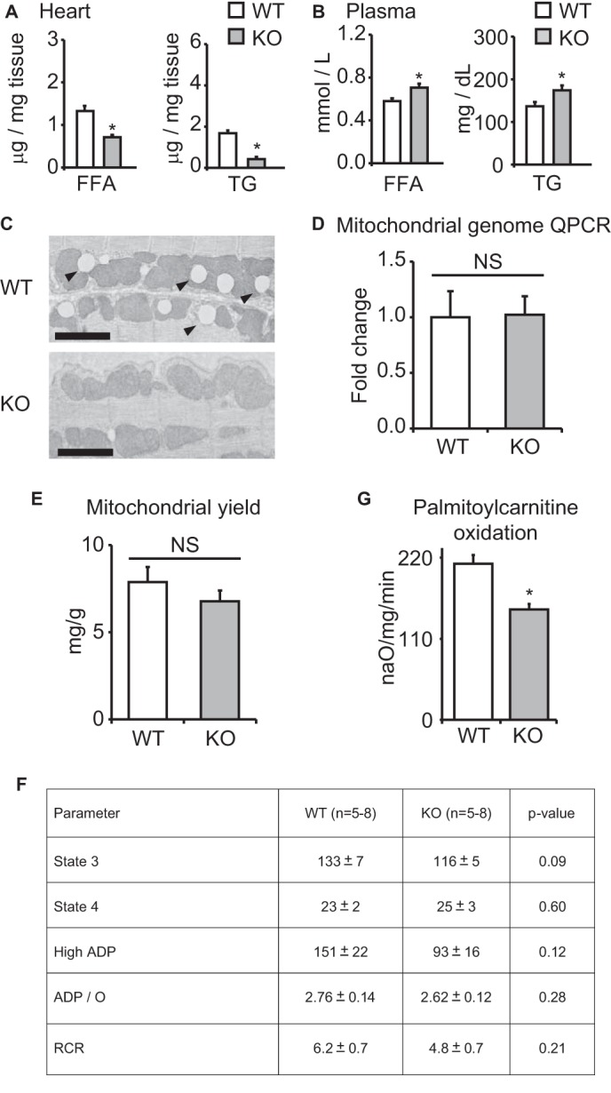
Mitochondrial parameters in KLF15 KO. Shown are FA and TG content in WT versus KO heart tissue (A) and plasma (B) (n = 5). *, p < 0.05 versus WT. C, representative transmission electron micrographs of WT versus KO heart tissue demonstrating marked paucity of intramyocellular lipid droplets (arrowheads) in KO tissue. Bar, 2 μm. D, qPCR for relative mitochondrial genome content in WT versus KO heart tissue (n = 4). Values are normalized to Rplp0 (36B4) genomic DNA. E, mitochondrial yield from whole heart tissue of WT versus KO mice, expressed as mg of mitochondrial protein/g of heart tissue (n = 4). F, rates of glutamate oxidation in mitochondria freshly isolated from WT versus KO hearts (n = 5–8). G, palmitoylcarnitine oxidation rates in freshly isolated mitochondria from WT versus KO hearts (n = 4). *, p < 0.05. Error bars, S.E.