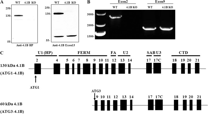
FIGURE 1.
Expression of 4.1B in MEF cells. A, immunoblot analysis of 4.1B expression in WT and 4.1B KO MEF cells. 40 μg of protein lysates were probed with polyclonal rabbit antibody against 4.1B head piece or 4.1B Exon 13. B, amplification of 4.1B cDNAs from MEF cells. The ATG1 or ATG3 4.1B transcripts were amplified as described under “Experimental Procedures.” Sizes of each PCR product, expressed in base pair (bp), are indicated. C, exon maps and protein structure of MEF cell 4.1B. Translation initiation sites are indicated (ATG-1, ATG-3). Domains are shown as described in the Introduction.

